Показать сокращенную информацию
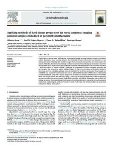
Applying methods of hard tissues preparation for wood anatomy: Imaging polished samples embedded in polymethylmethacrylate
| Автор | Arzac, Alberto | |
| Автор | López-Cepero, José M | |
| Автор | Babushkina, Elena | |
| Автор | Gomez, Santiago | |
| Дата внесения | 2019-07-01T07:22:00Z | |
| Дата, когда ресурс стал доступен | 2019-07-01T07:22:00Z | |
| Дата публикации | 2018-10 | |
| Библиографическое описание | Arzac, Alberto. Applying methods of hard tissues preparation for wood anatomy: Imaging polished samples embedded in polymethylmethacrylate [Текст] / Alberto Arzac, José M López-Cepero, Elena Babushkina, Santiago Gomez // Dendrochronologia. — 2018. — С. 76-81 | |
| ISSN | 11257865 | |
| URI (для ссылок/цитирований) | https://www.sciencedirect.com/science/article/pii/S1125786518301073 | |
| URI (для ссылок/цитирований) | https://elib.sfu-kras.ru/handle/2311/110553 | |
| Описание | Текст статьи не публикуется в открытом доступе в соответствии с политикой журнала. | |
| Аннотация | Cambial activity records short and long-term environmental signals in xylem anatomy, creating a permanent archive. Quantitative wood anatomy deciphers the relationship between cell structure and function in a spatiotemporal context. Obtaining high-resolution images of wood anatomical preparations is a critical stage in the process of decoding this information. Damage to cellular structures when sectioning by microtome is one of the main problems in the preparation of high-quality micro-sections. Cell damage leads to the occurrence of artifacts – most often related to broken cell walls – hindering the performance of image recognition programs, and increasing the time spent on the manual editing of images. In this work, we propose an alternative method to microtomy, based on embedding-polishing protocols established for hard tissue preparation. Wood samples are embedded in a transparent and non-reactive resin as polymethylmethacrylate (PMM) that is subsequently ground and polished. Being able to acquire images from the stained or unstained polished surfaces of the PMMblocks and sections (thinner than 100 μm) by using a wide range of optical methods such as reflected polarizing microscopy, epifluorescence microscopy, bright-field microscopy with diffuse illumination and circularly polarizing microscopy. This embedding method improves the mechanical integrity and quality of wood anatomical preparations, eliminating the problem of broken cell walls. Furthermore, this technique allows the preparation and analysis of large tissue surfaces. | |
| Тема | Embedding | |
| Тема | Microscopy | |
| Тема | Polymethylmethacrylate | |
| Тема | Surface staining | |
| Тема | Wood anatomy | |
| Название | Applying methods of hard tissues preparation for wood anatomy: Imaging polished samples embedded in polymethylmethacrylate | |
| Тип | Journal Article | |
| Тип | Published Journal Article | |
| Страницы | 76-81 | |
| ГРНТИ | 34.35.25 | |
| Дата обновления | 2019-07-01T07:22:00Z | |
| DOI | 10.1016/j.dendro.2018.08.005 | |
| Институт | Институт экологии и географии | |
| Институт | Хакасский технический институт — филиал СФУ | |
| Подразделение | Кафедра экологии и природопользования | |
| Подразделение | Кафедра строительства | |
| Журнал | Dendrochronologia | |
| Квартиль журнала в Scopus | Q1 | |
| Квартиль журнала в Web of Science | Q1 |